Brillouin spectroscopy is a challenging but powerful contact-free technique gathering rapidly growing interest in the biophysical community. Essentially, a pump laser is focused on a sample and the scattered light is spectrally resolved in the vicinity of the excitation wavelength. Upon interaction with spontaneous acoustic phonons, a small fraction of the photons are frequency shifted by an amount that depends on the mechanical properties of the material. The inherent challenges are; the extremely small spectral shifts, the overwhelming pump signal, and the very low signal amplitudes. LightMachinery has developed a VIPA-based Brillouin Hyperfine spectrometer and pump suppression module to optically probe the mechanical properties of biological (and non-biological) samples with unparalleled sensitivity. Its sub-picometer resolution, high contrast, strong pump suppression, and fast acquisition are indispensable for successful 2&3D Brillouin microspectroscopy or for monitoring real-time processes. The team of Prof. Dehoux and Prof. Margueritat recently performed a comparison study3 between the Lightmachinery system and the only other Brillouin spectrometer currently available commercially, a scanning multi-pass tandem Fabry-Pérot interferometers (TFPI). Their conclusions are published along with a fascinating investigation of the mechanical properties of multicellular tumor spheroids. “We have found similar performances [between the two Brillouin spectrometers] in terms of precision and accuracy, but shorter acquisition times for the VIPA-based technology for a given SNR. Both systems converge to a precision of ∼5 MHz for [the frequency shift] and ∼20MHz for [the linewidth]. However, the VIPA precision converges more rapidly, meaning that in the same experimental conditions, the VIPA-based spectrometer is about 50 times faster than the TFPI. (…) Given that the VIPA configuration is faster than TFPI under same experimental conditions, a crucial advantage for biological applications, we here use the VIPA spectrometer to probe the mechanics of multicellular tumor spheroids (MCTS).3”
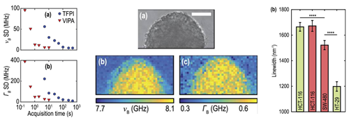
Figures obtained from [3]. Left:Standard deviation vs. acquisition time for Brillouin frequency shift (a) and linewidth (b). The HyperFine is ~50 times faster. Middle: Optical image (a) of a HCT-116 tumor spheroid and the corresponding maps of (b) the Brillouin frequency shift and (c) the Brillouin linewidth. Acquiring Brillouin maps is made possible by the high speed of the HyperFine system. Right: Brillouin linewidth of different colorectal cell lines. A significant difference is observed, demonstrating the ability to use this parameter to discriminate between cell lines.
[3]Yan, Guqi, et al. "Evaluation of commercial virtually imaged phase array and Fabry-Pérot based Brillouin spectrometers for applications to biology." Biomedical optics express 11.12 (2020): 6933-6944.


