The picometer resolution and other unique capabilities of the LightMachinery spectrometers open the possibility for exploring uncharted waters in various fields. We frequently collaborate with potential customers to test novel measurements and samples, or to fine tune the spectrometer design to facilitate specific applications. In many cases, these collaborations support researchers for grant applications (e.g. EPSRC, NSF, internal funding, etc) and provide them with a head start for their research. As an example, we collaborated with MSTATT LLC to measure the Brillouin signal of bovine enamel using the HF-8999-532-AUTO system7.
This work contributed to demonstrate the potential of using Brillouin spectroscopy for early cavity detection. In another study using the HF-8999-532-AUTO system, performed this time in collaboration with the CEA (Commissariat à l'énergie atomique et aux énergies alternatives, France), the suitability of VIPA-based Brillouin spectroscopy for Diamond Anvil Cell measurements was demonstrated8.
Currently, we are also collaborating directly with one of the largest microscopy companies in the world for developing a coupling module for the HyperFine systems. Overall, we have previous and ongoing collaborations on many other Brillouin projects as well as Raman spectroscopy, optical coherence tomography (OCT), and laser-induced breakdown spectroscopy projects (LIBS). We have collaborated or are currently collaborating with several companies in America, Europe, and Asia, as well as research institutes and universities including Sandia National Laboratories (USA), National Research Council Canada, Dutch Institute for Fundamental Energy Research (Netherlands), Institut National de la Recherche Scientific (Canada), Glasgow University (UK), University of Montreal (Canada), Drexel University (USA), University of Central California (USA), Leibniz University Hannover (Germany), Carleton University (Canada), Imperial London College (UK), etc.
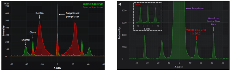
Figures obtained from [7] and [8]. Left: Brillouin spectra of bovine enamel (green) and dentin (red). Right: Brillouin frequency spectrum of water in a DAC at approximately 1 GPa. The inset shows the Brillouin spectrum of water at atmospheric pressure.
[7] Sophie Chagnon-Lessard, “Biological Brillouin Scattering with the Hyperfine Spectrometer.” 4th BioBrillouin Meeting, virtual, 2020.
[8] Sophie Chagnon-Lessard and David Walker. “Brillouin Spectroscopy in a Diamond Anvil Cell.” 2020, white paper, LightMachinery.


